Safe and Documented Grossing

Request a quote


Heavy duty casters
Adjustable height
Presence sensor
High power exhaust system
Transparent sliding cover
Twin tanks
All-in-one control panel
Display panel
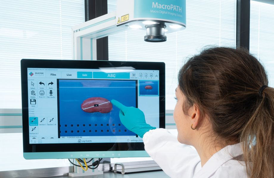
MacroPATH camera
MacroPATH PC
Foot pedals
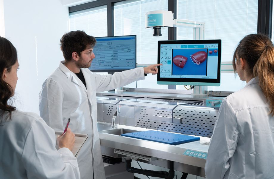
LIS monitor
Greater Ergonomics & Improved Flexibility
eGROSS is built around the user’s ergonomic needs, bringing a new level of comfort and efficiency. Its advanced features have been carefully designed to meet the specific needs of operators to achieve a seamless working experience. eGROSS offers a large working area made of corrosion-resistant stainless-steel surfaces, a dedicated formalin dispensing nozzle conveniently controlled by a foot pedal, and an easily accessible control panel with key functions to optimize the workflow.
A unique proximity sensor detects the presence of the operator. If no operator is detected for 5 minutes, the unit automatically switches off to extend the filter life. Installed on heavy duty casters, eGROSS can be easily moved without the need of a service engineer. The working height can be easily adjusted to accommodate standing or sitting according to operator’s habits.


Request a Virtual Demo

Enhanced User Safety
Macro Digital Documentation