Easy and Efficient Documentation of the Grossing Step
-
 Streamlined Workflow
 -
 Digitalize Grossing Documentation
 -
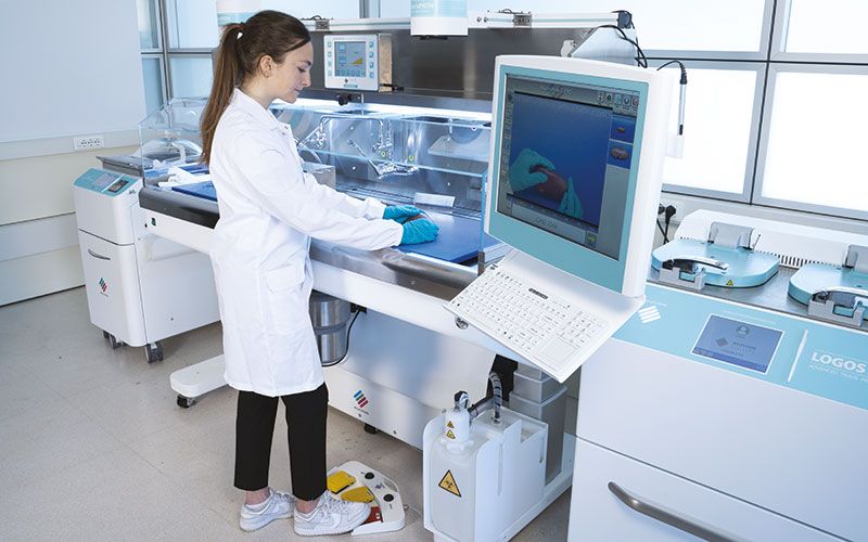
 Flexible Set-up
 -
 Full Integration with LIS & PACS
 -
 Enhanced Traceability
Request a quote

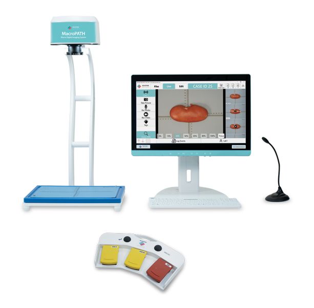
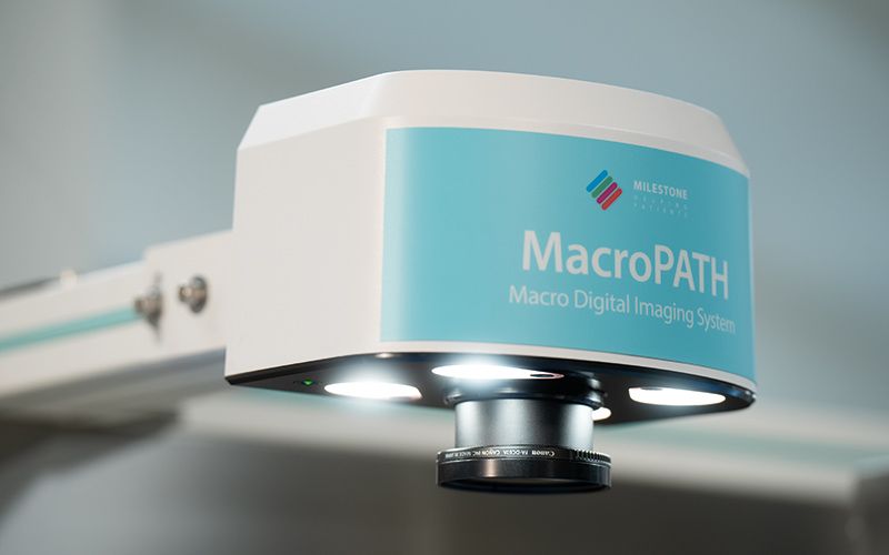
HIGH-RESOLUTION CAMERA
HIGH-PERFORMANCE TERMINAL
SMART USER INTERFACE
FOOT CONTROL
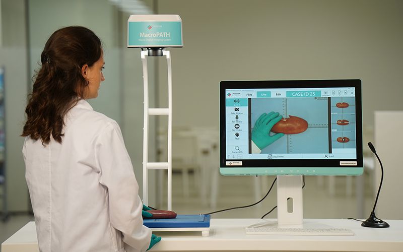
The sequence begins by entering the case ID through the barcode reader which automatically opens the case.
By using the built-in foot pedal or the voice control, the operator can easily zoom in and out, take images and record videos.
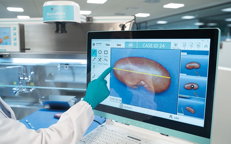
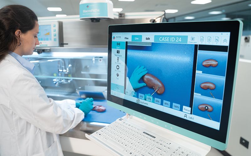
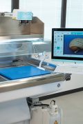
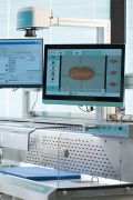
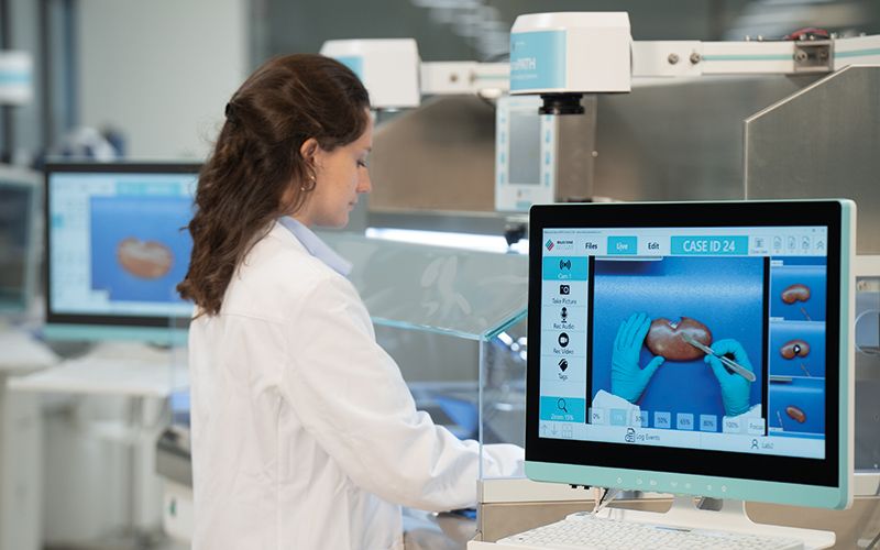
Digitalized Grossing Documentation
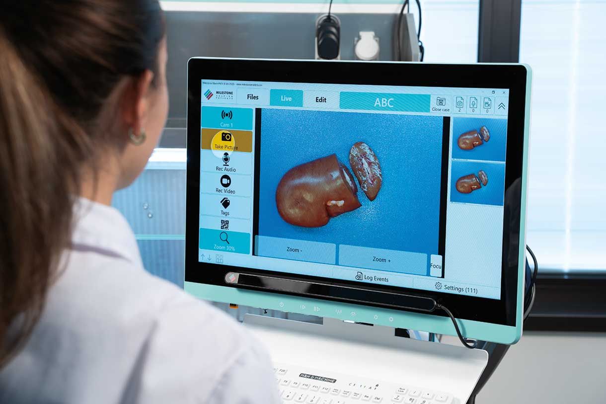
MacroPATH is a comprehensive tool designed to provide digital documentation of the grossing of surgical specimens and biopsies.
It is equipped with a high-resolution built-in camera and microphone, enabling the capture of images, videos and audios of the biospecimen and its dissection in exceptional detail. It also includes an advanced suite of tools for image sizing and editing, along with the capability to generate comprehensive reports.
MacroPATH is an all-in-one platform for integrated and efficient documentation of the grossing process.
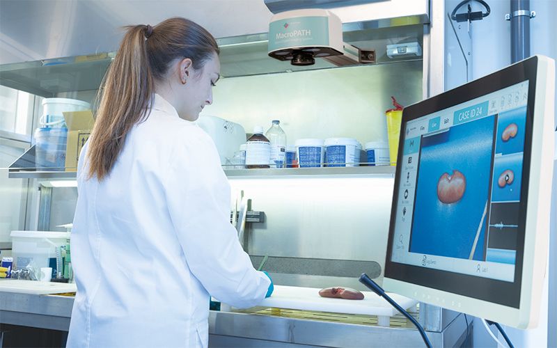
Flexible Set-up
MacroPATH offers flexible installation options to suit the needs of different labs. It can be used as a stand-alone system or integrated as a complementary component into any grossing station. Its versatility perfectly aligns with the modularity of the Milestone grossing station, UltraGROSS, allowing for the installation of the MacroPATH touchscreen terminal in either frontal or lateral positions, based on the specific requirements and preferences of the operator.
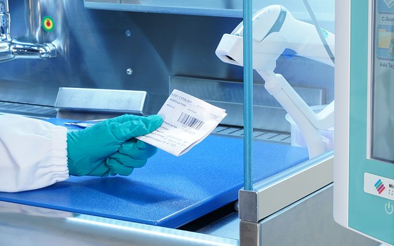
Enhanced Traceability
Integration with LIS & PACS
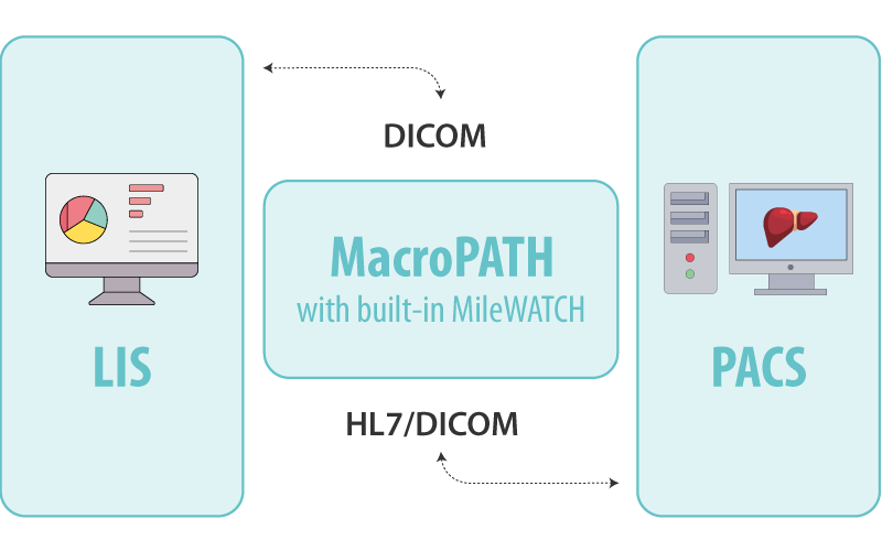
As a result, MacroPATH, thanks to the built-in MileWATCH, is integrated with LIS and PACS through various protocols, including DICOM, HL7, TWAIN and WIA, ensuring the highest level of compliance with key standards.
MileWATCH
24/7 Tracking and Monitoring
Track, monitor and manage specimens throughout their journey.
Discover More
The Perfect Partners to your MacroPATH
C-Scan
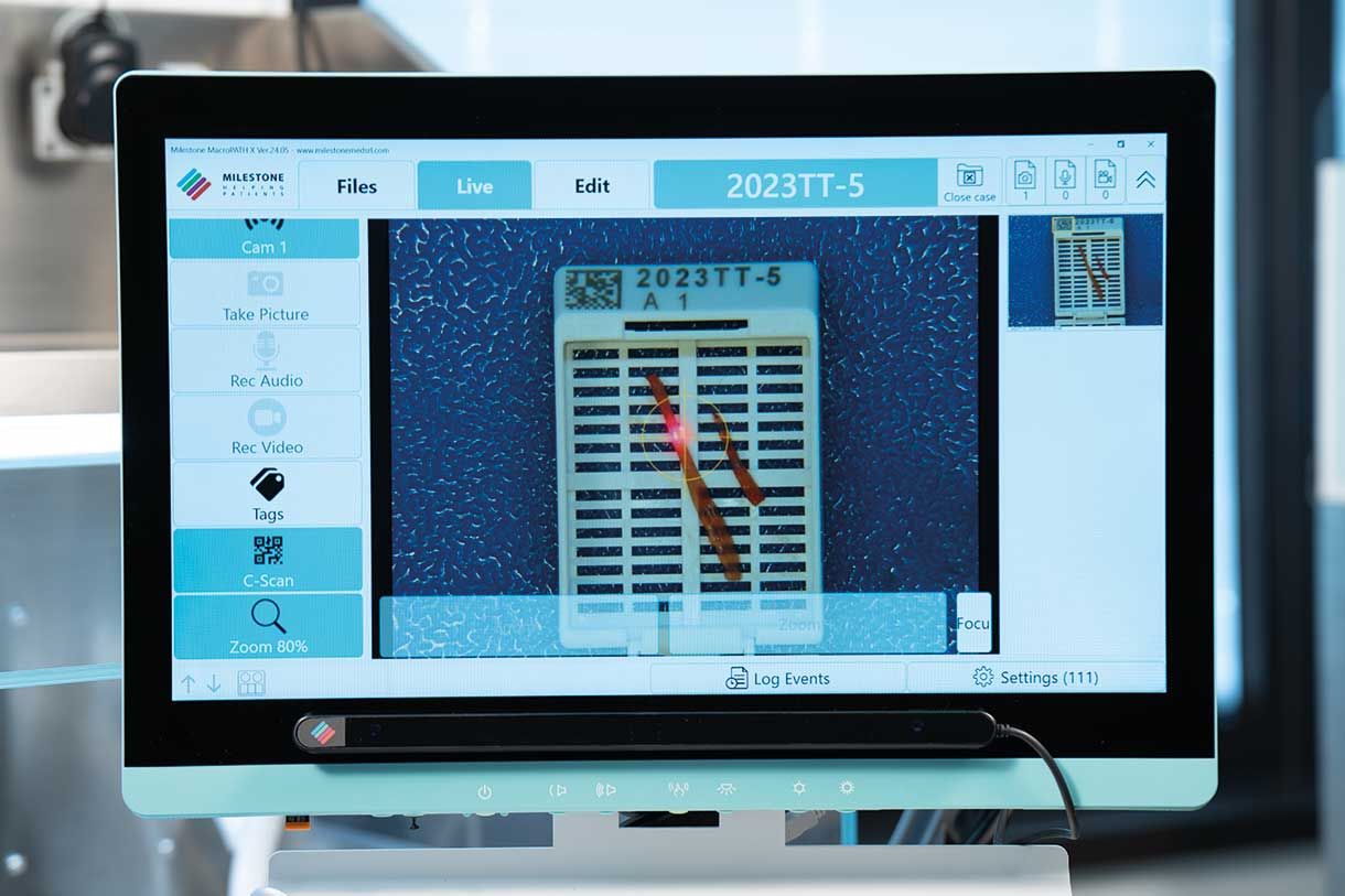
C-Scan enables effortless operation as it is ready to use. The operator simply places the cassette with biopsy under the laser beam. C-Scan automatically reads the cassette’s code, opens the case ID folder and takes a picture at a preset zoom.
LOOX
The LOOX advanced IR sensor bar tracks the operator’s eye movement. The operator can smoothly perform tasks such as zooming, capturing images, and recording videos or audios by simply looking at the command icons on the screen. This allows for hands-free documentation during the grossing phase.