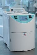
Pioneering Fixation & Tissue Processing
MAGNUS redefines tissue processing with its innovative dual retort system, enabling continuous batch loading without the need for time-consuming cleaning cycles. This unique design delivers unmatched throughput and shorter processing times, allowing extended fixation for superior formalin penetration and exceptional tissue quality.
-
 Superior Turn-Around Time
 -
High Quality Results
-
Ease of Use – No Reagents Transfer
-
Specimen and Personnel Safety
-
Cost and Time Savings
Request a quote

ENCLOSED CABINET
DUAL ROBOTIC ARM
INDEPENDENT LOADING RETORT
INDEPENDENT WAX RETORT
AUXILIARY WAX RESERVOIR
ICON-DRIVEN INTERFACE
EASY ACCESS
SLIDING DRAWER
HIGHLY-SENSITIVE ULTRASONIC SENSOR


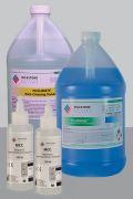
Effortless Operation with No Reagent Transfer
MAGNUS simplifies reagent management by using commercially available jugs, eliminating the need for decanting. Operators can replace exhausted jugs with new ones in seconds, streamlining the process and enhancing safety. Key benefits include:
- No Decanting: Direct use of jugs saves time and reduces handling.
- Zero Fume Exposure: Sealed reagent changes protect operators from toxic fumes.
- Time Efficiency: Eliminates cleanup of spills, freeing up technician time.
- Error-Free Operation: Barcode scanning ensures accurate reagent placement, preventing errors.
MAGNUS delivers unmatched ease of use, safety, and efficiency for seamless laboratory workflows.

Significant Cost and Time Savings
MAGNUS optimizes laboratory efficiency by reducing costs and turnaround time (TAT). By eliminating xylene and clearing solvents, MAGNUS lowers toxic reagent disposal costs and extends paraffin wax life up to a month. The dedicated wax retort eliminates pump-in/out processes and cleaning cycles, allowing immediate start of new processing runs, saving reagents and improving TAT. Additionally, the removal of reagent decanting minimizes downtime, further enhancing productivity.
MAGNUS delivers substantial savings and streamlined workflows for cost-effective, high-performance tissue processing.
MileWATCH
24/7 Tracking and Monitoring
Track, monitor and manage specimens throughout their journey.
Discover More
The Perfect Partners to your MAGNUS
Auto-Embedding with Synergy